輔助診斷臨床應用型 AI 及人工智慧模型自行建立平台
  |
我們的解決方案,包含三大領域,並可相互整合:
一、Aiforia Clinical Suite 人工智慧臨床診斷模組
- 具備 CE-IVD 認證,無須重新進行訓練開發,可直接使用於臨床診斷。
- 取得品質管理系統。
- Certified Quality Management System(ISO 13485)及資訊安全管理系統認證(ISO 27001)。
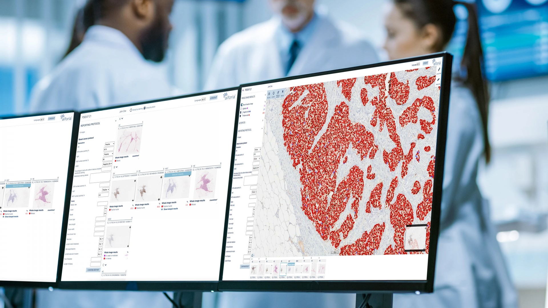
- 僅需將玻片上傳至雲端平台,不需假他人之手,便可輕鬆建立自己的 AI 模型。
- 利用此平台,我們可為您提供使用諮詢及教育訓練服務,後續的訓練及模型建立則可以由客戶自由進行,研究進度、概念及研究成果均掌握在客戶之手。
若您有意將您的模型轉化為商業模型,我們亦可提供此類協助。
三、Education Hub 雲端教學平台- 建立於雲端之教學平台,可提供玻片教育、閱片分享、雲端資料儲存及遠端會診功能。


 |
|||||||
 |
已有超過 2 百萬張影像使用 Aiforia 平台進行分析 | 
|
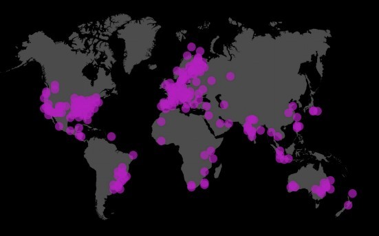
全球超過 5,000 個用戶,其中包含全球的病理學家、醫師及研究人員使用 Aiforia 的 AI 及影像分析工具 |  |
已開發過超過 400 個以上用於影像分析的 AI 模型,超過 60 篇以上國際期合作發表刊 | 
等平台進行整合
|
支援所有主要玻片掃描儀及顯微影像系統及 DICOM 格式,並可與 LIS、PACS 等平台進行整合 |
一、Aiforia Clinical Suite 人工智慧臨床診斷模組
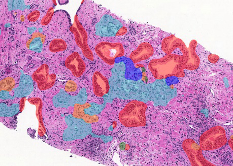
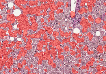
以 AI 工具輔助病理醫師提高診斷效率、降低瑣碎的量化分析工作、強化判讀精準度乳癌 Aiforia® Clinical AI Model for Breast Cancer; ER |
乳癌 Aiforia® Clinical AI Model for Breast Cancer; PR |
乳癌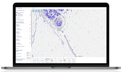 Aiforia® Clinical AI Model for Breast Cancer; Ki-67 |
前列腺癌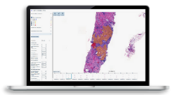 Aiforia® Clinical AI Model for Prostate Cancer; Gleason Grade Groups |
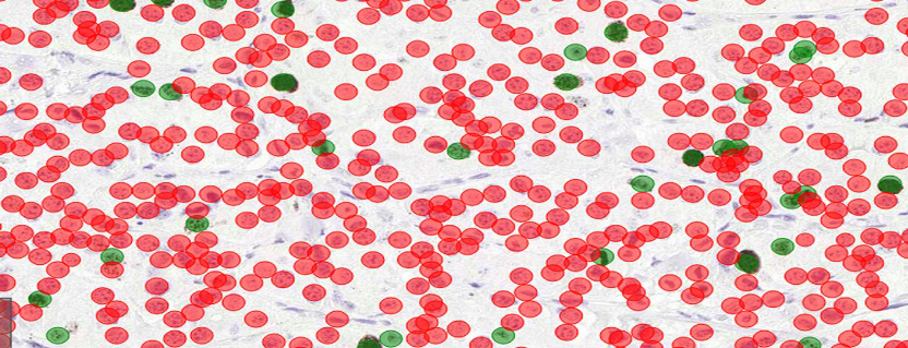
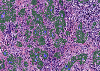
肺癌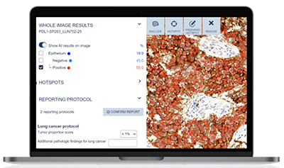 Aiforia® Clinical AI Model for Lung Cancer; PD-L1 |
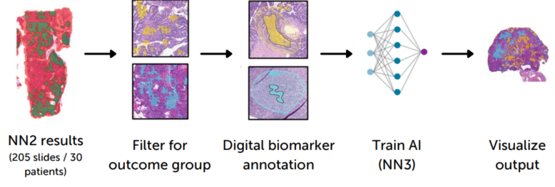
二、Aiforia Create人工智慧自建模組
AI 應用於研究領域
- 可為任何 2D 影像(如病理組織切片、放射科影像)、各種影像格式(各家掃片機、顯微鏡影像系統、DICOM、他牌 AI 平台輸出影像格式)建立深度學習 AI 模型,進行定量分析。
- 可為使用者量身打造 AI 模型 / 客制化服務。
- 超過 60 篇以期刊論文,發表於神經科學領域、腫瘤 / 癌症領域及肝臟或其他醫學領域。
模型範例:
 |
  |
|
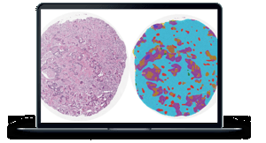
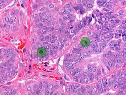
Ki-67 quantification |
PD-L1 scoring |
 |
 |
|
Tumor outcome prediction |
Carcinoma segmentation |
  |
 |
|
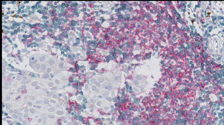
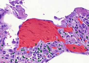
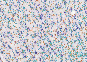
HER2+ marker detection AI assisted detection of IHC stained CD3+ T-cells, CD20+ B-cells, DC-Lamp+ mature dendritic cells, and Hematoxylin in a HER2+ breast cancer surgical resection at the Royal College of Surgeons in Ireland. |
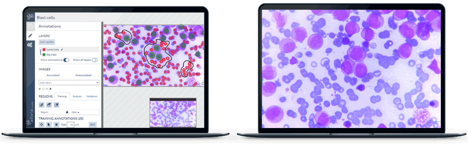
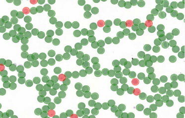
Blast cell and RBC identification AI trained to identify blast cells and red blood cells on previously diagnosed cases of acute myelogenous leukemia at the Jinnah Sindh Medical University. |
 |
 |
 |
|
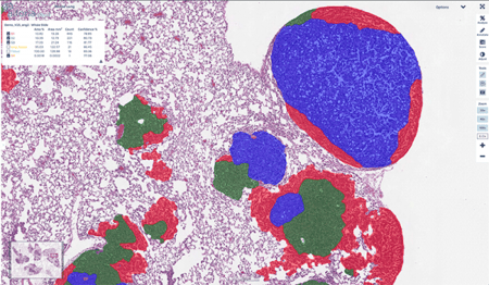
Tumor grading Automating tumor grading in non-small cell lung cancer(NSCLC)studies at MIT. |
Tumor grading Automated tumor grading in prostate cancer biopsies at the Helsinki University Hospital, Finland, and in Region Skåne lab, Sweden. |
Tumor grading Automated tumor grading in breast cancer biopsies. |
 |
  |
|
Mitosis Mitosis detection and quantification. |
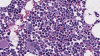
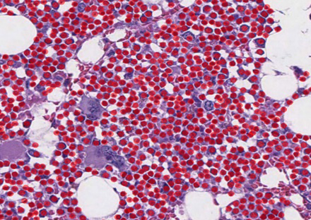
Hematopoietic cell enumeration*Bone Marrow AI model used to identify and enumerate hematopoietic cells to determine changes in bone marrow cellularity for pharmacologic safety studies at Charles River Laboratories. |
  |
  |
|
Malaria detection Automated malaria parasite detection from blood smears at the University of Helsinki. |
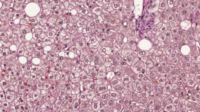
Ballooning assessment AI in studying nonalcoholic fatty liver disease(NAFLD)and its capability to segment structures in liver histology at the University of Helsinki. |
  |
  |
|
Fibrosis quantification Automated quantification of damage and scarring in liver tissue in nonalcoholic fatty liver disease(NAFLD)and nonalcoholic steatohepatitis(NASH). |
Pulmonary fibrosis quantification Identification and quantification of histopathological features of idiopathic pulmonary fibrosis(IPF)including fibroblast foci, and interstitial and alveolar inflammatory cells at the University of Helsinki. |
  |
  |
|
Tuberculosis quantification Automated detection and quantification of cells and lesions in tuberculosis studies at Tufts University. |
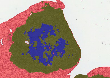
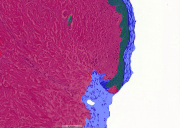
Dermis and epidermis detection Automated detection of dermis and epidermis of skin tissue sample. |
  |
  |
|
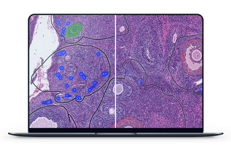
Follicle quantification Preclinical studies on identification and quantification of the different stages of ovarian follicles in rats using AI models at Charles River Laboratories. |
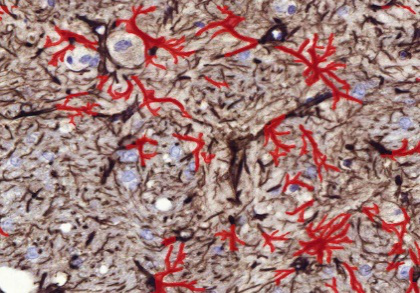
Astrocyte reactivity quantification Quantifying astrocyte reactivity in preclinical neurotoxicity studies at Orion pharmaceutical company. |
  |
  |
|
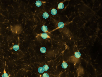
Astrocyte detection Use of AI for image analysis and astrocyte detection in Parkinson’s Disease research at Vall d'Hebron Instituto de Investigación. |
Amyloid-β detection AI-assisted identification of the most characteristic markers of AD and CAA, including Aβ plaques, at Massachusetts General Hospital. |
  |
  |
|
α-synuclein assessment Quantitative assessment of alpha-synuclein pathology in preclinical Parkinson's disease studies at Lundbeck pharmaceutical company. |
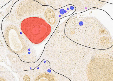
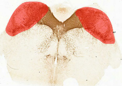
Nigra area detection Automated nigra area detection. |
  |
  |
|
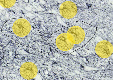
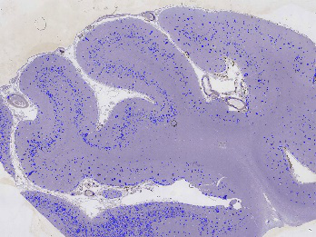
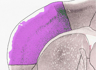
Whole slide human brain segmentation Grey/White matter segmentation and Neuron/Glia counts in human brain sample. |
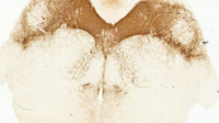
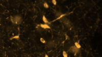
TH+ neuron quantification Automated dopaminergic neuron quantification in Parkinson’s disease research at Duke University. |
三、Education Hub 雲端教學平台
- 此平台是專為組織學、解剖學、病理學、寄生蟲學及獸醫病理學等課程所設計的。
- 可以使老師及學生均不再受限於顯微鏡、固定的教室及書面資料。
- 雲端平台可以接受老師使用任何玻片掃描儀所上傳的任何影像,並直接與學生分享。
- 老師可以於平台中加入影像的註解、說明及影像的病史、個案背景訊息等,完整整個樣本的起源及狀態描述,豐富學生的學習資源。
- 此平台基於軟體即服務 SaaS(Software-as-a-Service)模式,可以由簡單的研討會模式擴展延伸至大型進階版的教學模式。
合作夥伴             |