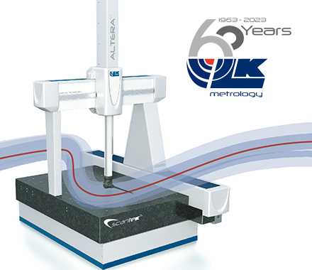

Metafer 自動化細胞掃描分析工作站
● 搭配高精確電動載物台,可一次掃瞄八張玻片
● 同一硬體平台上,可擴充MSearch分裂中期細胞掃描儀、MetaCyte間期細胞螢光點掃描儀、RCDetect罕有細胞自動掃描儀及CometScan單細胞凝膠電泳分析儀
● MSearch: 可自動在亮場或螢光圖像中辨識分裂中期細胞染色體及紀錄座標
● MetaCyte: 可同時檢測六種螢光染料,自動紀錄每個細胞中的探針標定數目並製成統計表格
● RCDetect 根據螢光強度、色彩、細胞形狀,自動進行高通量的特定罕有細胞搜尋,如骨髓細胞樣本中的神經母細胞瘤
● 同一硬體平台上,可擴充MSearch分裂中期細胞掃描儀、MetaCyte間期細胞螢光點掃描儀、RCDetect罕有細胞自動掃描儀及CometScan單細胞凝膠電泳分析儀
● MSearch: 可自動在亮場或螢光圖像中辨識分裂中期細胞染色體及紀錄座標
● MetaCyte: 可同時檢測六種螢光染料,自動紀錄每個細胞中的探針標定數目並製成統計表格
● RCDetect 根據螢光強度、色彩、細胞形狀,自動進行高通量的特定罕有細胞搜尋,如骨髓細胞樣本中的神經母細胞瘤
Sequential analysis of the same neuroblastoma cell with GD2 specific antibody and KI-67 proliferation marker (left), TUNEL assay (center) and FISH (right) showing a trisomy of chromosome #1 in the GD2-positive cell (FISH signals: red = D1Z1 [1p36]; green = D1Z1 [centromere 1]). Image courtesy of G. Mehes, University of Pecs, Hungary.
Applications
Interphase FISH Signal Analysis (Spot Counting)
Detection of Gene Fusions
Detection of Gene Amplification in Tissue Sections
Microwell Based Cell Screening
原廠連結: